Browse Articles
Conditioning Medicine
International bi-monthly journal of cell signaling, tissue protection, and translational research.
Current Location:Home / Browse Articles
Commentary: Can astrocytic mitochondria therapy be used as antioxidant conditioning to protect neurons?
Time:2023-04-22
Number:7227
Kazuhide Hayakawa1
Author Affiliations


- 1Neuroprotection Research Laboratories, Departments of Radiology and Neurology, Massachusetts General Hospital and Harvard Medical School, Charlestown, Massachusetts, USA.
Conditioning Medicine 2022. 5(6): 192-195.
Abstract
In the context of central nervous system (CNS) disease, oxidative stress may cause progression of cell death and neuroinflammation. Therefore, restoring mitochondrial antioxidant ability within cells is a major therapeutic strategy in many CNS disorders. A recent study uncovers a novel mechanism of astrocytic mitochondria being neuroprotective after intracerebral hemorrhage in mice. In their work, systemic administration of mitochondria obtained from astrocytes restores neuronal antioxidant defense, prevents neuronal death while promoting neurite outgrowth, indicating that extracellular mitochondria may play key roles in mediating beneficial non-cell autonomous effects. Given that mitochondria are also responsible for tolerance to stress and injury, is it possible that exogenous mitochondria signals may regulate cellular conditioning by boosting antioxidant ability? Further studies are warranted to build on these emerging findings in the pursuit of conditioning therapies mediated by mitochondrial transplantation in CNS injury and disease.
Keywords: Astrocytic mitochondria, Mitochondrial transplantation, Antioxidant defense, CNS disorders
Mitochondria are the intracellular energetic core of cells and they are essential for maintaining cellular function in mammals (Devine and Kittler, 2018). The brain is a high-metabolism organ wherein mitochondria play a central role in almost all aspects of cellular homeostasis during pre- and post-conditioning (Bastian et al., 2018; Narayanan and Perez-Pinzon, 2017; Simon, 2014; Zerimech et al., 2021), aging (Selvaraji et al., 2019) and many central nervous system (CNS) disorders including stroke (Rutkai et al., 2019; Wang et al., 2018). In the context of injury or disease in the CNS, accumulating mitochondrial ROS and inflammasome, along with imbalanced mitochondrial membrane permeability, activates cell death pathway and neuroinflammation (Green, et al., 2011). Therefore, restoring mitochondrial perturbation within cells is a major therapeutic strategy (Golanov, 2019).
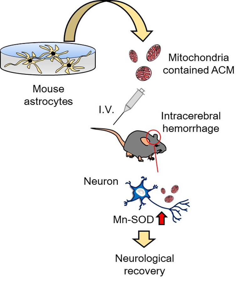
In the context of a damaged and recovering brain, crosstalk between neuronal, glial, and vascular cells within neurovascular units may support neuroplasticity through the intercellular exchange of non-cell autonomous signals so called “help-me signaling” including small peptides, microRNAs, and extracellular vesicles (Esposito, 2018; Salmeron, 2018; Lopez et al., 2017; Xing and Lo, 2016). Recently, it has been demonstrated that mitochondria can also be secreted, and extracellular mitochondria play key roles in CNS disease (Hayakawa et al., 2018). Injured neurons may release damaged mitochondria that transfer to astrocytes for transmitophagy (Davis et al., 2014), while mitochondria may also be transferred from astrocytes into adjacent neurons in a protective manner (Hayakawa et al., 2016). Because extracellular mitochondria are incorporated into cells, exogenous mitochondria transplantation has been pursued as a candidate therapeutic approach for CNS injury and disease (Chen et al., 2020; Chou et al., 2017; Nakamura et al., 2019). In a proof-of-concept study in the heart, McCully and colleagues (Emani et al., 2017) demonstrated that direct transplantation of autologous mitochondria isolated from the patient’s skeletal muscle into the heart was feasible without inducing immune or auto-immune responses. In animal models of regional ischemia-reperfusion injury, intracoronary injection of autologous mitochondria decreased infarct size, enhance coronary blood flow, and increase cardiac function (Blitzer et al., 2020; Guariento et al., 2020; Shin et al., 2019), suggesting that mitochondrial transplantation therapy may be applicable in other injuries or diseases. A recent paper by Tashiro et al (2022) has now expanded upon the concept of therapeutic use of extracellular mitochondria to show that systemic administration of astrocyte-originated mitochondria boosts neuronal antioxidant defense and promotes recovery after intracerebral hemorrhage (ICH) in mice (Figure 1).

In a new window | Download PPT
Figure 1: Potential mitochondria therapy by treatment with conditioned media collected from cultured astrocytes.
In the study, the authors first showed that after the injury onset, mitochondrial anti-oxidative defense system was disrupted along with worsening oxidative damage in the affected brain hemisphere. Immunohistochemistry, western blot, and qPCR analysis demonstrated that the manganese superoxide dismutase (Mn-SOD) expression was significantly decreased in the ipsilateral hemisphere while superoxide radicals and nitric oxide such as O2.- production and 3-nitro-tyrosine formation were robustly increased compared with the contralateral hemisphere. The authors attempted to reverse the oxidative stress responses through administration of astrocyte-derived mitochondria. The mitochondria of astrocytes in culture were fluorescently labeled with MitoTracker-Red/CMXRos, and the extracellularly released labeled mitochondria were collected and intravenously injected 24 h after the injury. Notably, approximately 62.9% of neurons in the ipsilateral hemisphere appeared to uptake infused mitochondria 24 h after injection. Given that mitochondrial incorporation in the cardiomyocytes can be around 23% following intracoronary infusion (Cowan et al., 2016), the exciting finding of the efficient mitochondria uptake in neurons following intravenous administration may lead to the opportunity to investigate a quantitative parameter of how many mitochondria are required to treat the number of damaged neurons. Moreover, intravenous infusion may impact other organs besides the CNS. Therefore, future studies are warranted to investigate CNS-targeting delivery for clinical translation.
Next, the authors examined whether administration of astrocytic mitochondria can improve outcomes after the injury. The loss of function of astrocytic mitochondria was conducted by confirming that 78% of mitochondria from astrocyte culture media (ACM) were removed by 0.22 μm filtration, consistent with previous studies (Hayakawa et al., 2016; Jung et al., 2020). Then, ACM or mitochondria depleted ACM (mdACM) were administered once a week for three weeks after ICH followed by assessing neurological deficit sore (NDS). The authors demonstrated that the NDS score was significantly reduced in ACM-treated mice at day 21, whereas mdACM treatment did not improve NDS score. Interestingly, ACM-treated mice showed significant restoration of Mn-SOD in the neurons throughout peri-hematomal brain areas, but again mdACM did not show restoration of Mn-SOD expression in neurons. It has been reported that intracellular Mn-SOD mediated neuroprotection correlates with reduced ROS overproduction, decreased neuronal death, and improved neurologic deficits after ischemic injury (Murakami et al., 1998). But are these beneficial mechanisms regulated by non-cell autonomously?
An important question is whether astrocytic mitochondria directly or indirectly provide Mn-SOD to the damaged neurons. To address the question, the authors rigorously conducted loss-of-function assays in vitro by mitochondrial removal from ACM, Mn-SOD siRNA transfection, or using cultured primary astrocytes from GFAPCreERT2:Mn-SODfl/fl mice. Initially, the authors confirmed that ACM protected neurons exposed to red blood cell lysate mimicking ICH-like injury, which was accompanied by reduced ROS production and increased expressions of Mn-SOD protein and mRNA. When Mn-SOD was suppressed in astrocytes by either siRNA or Mn-SOD knockout, the ACM-mediated antioxidant defense was clearly diminished, suggesting that astrocytic mitochondria may directly provide their Mn-SOD to neurons to counteract oxidative stress. Mn-SOD is vital to the protection of cells and is induced by both pre- and post-conditioning (Danielisova et al., 2006; Yamashita et al., 1994). This raises an intriguing question whether extracellular mitochondria signals may increase cellular tolerance to stress and injury by boosting antioxidant ability.
Finally, the authors designed experiments to investigate whether the previously found mitochondria-encoded peptide humanin (HN) (Jung et al., 2020) regulated the expression of Mn-SOD, anti-oxidative defense, and neurite outgrowth in damaged neurons. Intriguingly, treatment with HN in injured neurons inhibited ROS generation and protected cells from oxidative stress-induced cell death by activation of STAT3 (signal transducer and activator of transcription 3) along with upregulating Mn-SOD expression and preventing ROS overproduction, suggesting that the mitochondria-derived small peptide HN may also be one of the key effectors in astrocytic mitochondrial transfer-mediated neuroprotection. The authors also noted that they found that extracellular mitochondria from injured astrocytes in vitro had lower functionality compared to the control group. It is critical to consider that the beneficiality of mitochondrial transfer may depend on context. For instance, activated astrocytes after focal cerebral ischemia may express regenerative genes, whereas neurodegenerative diseases activate the proinflammatory phenotype and induce an inflammatory microenvironment. In this context, proinflammatory astrocytes are enriched in addition to proinflammatory microglia that exacerbates neurodegeneration through intercellular crosstalk via fragmented mitochondria (Joshi et al., 2019).
Tashiro et al (2022) has uncovered a new mechanism for astrocytic mitochondria-mediated neuroprotection through boosting antioxidant defense mediated by Mn-SOD in damaged neurons in the brain. These exciting findings reveal the feasibility of therapeutic mitochondrial transplantation potentially “conditioning” cells by amplifying the ability to counteract oxidative stress. It has been reported that mouse mitochondria efficiently fused to the mitochondrial network in human cells, while nuclear and mitochondrial genomes may need to be the same to retain functional compatibility with one another (Yoon et al., 2007). Future research that investigates a quantitative threshold, quality control, and the effect of different species of astrocyte-derived mitochondria may lead to the novel strategy of antioxidant pre- or post-conditioning via therapeutic mitochondrial transplantation in stroke and other CNS disorders.
Competing Interests
The authors declare they have no competing financial interest.
References
Kazuhide Hayakawa
Neuroprotection Research Laboratories, Departments of Radiology and Neurology, Massachusetts General Hospital and Harvard Medical School, Charlestown, Massachusetts, USA.
Corresponding author:
Kazuhide Hayakawa
Email: khayakawa1@mgh.harvard.edu

In a new window | Download PPT
Figure 1: Potential mitochondria therapy by treatment with conditioned media collected from cultured astrocytes.
Supporting Information
Metrics
| Full-Text | Supporting Information | ||
|---|---|---|---|
| Number | 7227 | 13 | 0 |
Copyright © 2017 Conditioning Medicine, All Rights Reserved.
Address: Conditioning Medicine Editorial Office, 3500 Terrace Street, Pittsburgh, PA, 15213, USA